Case Report | | Peer-Reviewed

Effects of Low-Load Exercises with Blood Flow Restriction on Shoulder of a Multiple Sclerosis Patient with Shoulder Tendinopathy: A Case Study

Received: 2 July 2026     Accepted: 20 July 2026     Published: 17 August 2026
Views:       Downloads:
Abstract

Blood flow restriction (BFR) training involves placing an inflatable cuff around the proximal part of a limb to partially restrict arterial inflow during exercise. When combined with low-load resistance exercise (LLRE), this approach has been shown to produce improvements in muscle strength and hypertrophy comparable to those achieved with conventional high-load resistance training in healthy individuals. Through its proximal effects, LLRE combined with BFR may also enhance muscle strength and size in individuals with supraspinatus tendinopathy. Although high-load resistance training is effective for increasing muscle strength and hypertrophy, its application in people with multiple sclerosis (MS) is often limited because of fatigue. This case report describes the proximal effects of an LLRE+BFR program in a patient with MS and supraspinatus tendinopathy. Our case was a 48-year-old woman with a 5-year history of relapsing-remitting MS, who presented with right shoulder pain and muscle weakness in the right shoulder at rest and while elevation. She received a LLRE+BFR program for her right shoulder and aerobic exercises based on the BFR protocols in 12 sessions over 4 weeks. Outcome measures were collected before the intervention and after 4 weeks and included: 1) strength of supraspinatus and four scapular stabilizer muscles. 2) thickness of supraspinatus and upper trapezius muscles. 3) thickness of supraspinatus tendon 4) pain pressure threshold of supraspinatus and deltoid muscles. 5) Supraspinatus muscle echogenicity. Improvement was observed in the strength of supraspinatus and scapular stabilizers. Thickness of supraspinatus and upper trapezius muscles and pain pressure threshold in the supraspinatus and deltoid muscles also increased. Furthermore, supraspinatus echogenicity and tendon thickness decreased. The patient experienced a significant improvement in all outcomes following the use of a LLRE+BFR program. This program may be an option for strengthening training with less stress in MS people with supraspinatus tendinopathy.

Published in International Journal of Sports Science and Physical Education (Volume 11, Issue 2)
DOI 10.11648/j.ijsspe.20261102.12
Page(s) 32-44
Creative Commons

This is an Open Access article, distributed under the terms of the Creative Commons Attribution 4.0 International License (http://creativecommons.org/licenses/by/4.0/), which permits unrestricted use, distribution and reproduction in any medium or format, provided the original work is properly cited.

Copyright

Copyright © The Author(s), 2026. Published by Science Publishing Group

Keywords

Blood Flow Restriction, Proximal Effect, Supraspinatus Tendinopathy, Multiple Sclerosis

1. Introduction
Shoulder pain affects up to half of the population annually , with rotator cuff tendinopathy as one of the most common causes, accounting for 30 to 70 percent of shoulder pain cases . Previous studies have shown that exercise yields promising results in managing rotator cuff tendinopathy . High-load resistance exercises (HLRE), which uses external loadings of 60-90% of 1-repetition maximum) are the golden standard for enhancing muscle strength and inducing hypertrophy, but such intense loads may not be suitable for frail populations .
A promising alternative for strength training is blood flow restriction (BFR), which can achieve similar outcomes to high-load training by using less exercise load and tissue stress . BFR includes a cuff encircling the proximal part of the upper or lower limb, filled with air. The cuff restricts blood flow and increases pressure on the extremity, resulting in decreased arterial blood circulation and cessation of venous return . In recent years, studies have shown that low-load resistance exercises (20-30% of 1 RM) combined with BFR (LLRE+BFR) can increase muscle strength and hypertrophy .
A 2017 systematic review by Hughes and Lixandrao suggested that LLRE+BFR can induce muscle hypertrophy comparable to HLRE, but the improvement in muscle strength following LLRE+BFR is comparatively lower than that achieved with HLRE . A systematic review published in 2020 reported that LLRE combined with BFR not only minimizes mechanical stress on the tissues but also produces improvements in muscle strength and hypertrophy comparable to those achieved with HLRE . In addition, gains in muscle size and strength may occur sooner with LLRE+BFR than with HLRE . Moreover, several studies have investigated the pain-relieving effects of BFR-assisted exercise in patients with different musculoskeletal conditions . Therefore, the application of LLRE+BFR can be a potential alternative to increase muscle strength and hypertrophy while alleviating pain in patients with musculoskeletal injuries. This technique is particularly suitable for the elderly and individuals with conditions like Multiple Sclerosis (MS), who may have lower tolerance for high loads.
MS is a central nervous system disease that affects approximately 2.2 million people worldwide annually . Progressive weakness and motor fatigability are two main complaints among most people with MS, which can contribute to other problems such as reduced quality of life and performance . Strength training has many advantages for MS patients , however they may perform lower doses which accompanies less improvement . This is because many people with MS may experience motor fatigability and temporary increase in sensory symptoms during exercise . Recent reviews strengthened the evidence supporting BFR in neurological conditions. These studies report that BFR can improve muscle strength, functional performance, muscle mass, and pain across several neurological disorders, including MS .
In this study, we examined an MS patient suffering from rotator cuff tendinopathy. LLRE+BFR is potentially effective in this case as it can increase strength and alleviate pain without inducing motor fatigability . Hence, the aim of this study is to evaluate the proximal effects of combining LLRE and BFR on the strength of supraspinatus and stabilizers of scapula muscles, thickness, and echogenicity of supraspinatus muscle, and pain on an MS patient with supraspinatus tendinopathy.
2. Case Description
The patient was a 48-year-old woman with a history of Relapsing-Remitting MS for about 5 years. She was in the remitting phase during the treatment, and was prescribed Rituximab by her neurologist. The most disabling MS symptom was fatigue, which was aggravated by moderate to even low levels of activity. Fatigue Severity Scale (FSS) was used to measure fatigue. It consists of 9 items, each scored on a 7-point Likert scale, where higher scores representing greater fatigue.
The main complaint of the patient to come under the care of this team was her shoulder pain at rest and while movement unrelated to MS, which had been started a month before and was not related to her MS. She was also suffering from night pain which caused sleep disturbance. The patient was not athletic and was prone to fatigability if prescribed with a high-load training regimen. Hence the team discussed the potential benefits of using BFR+LLRE to improve her condition, while not exacerbating her MS symptoms. An informed consent was received to publish the information of this case report.
2.1. Physical Therapist Examination
She performed a full passive range of motion with no pain; however, active abduction and flexion were restricted to 90 degrees though also painless, suggesting weakness in the shoulder muscles. Following a thorough history-taking, specific tests including Hawkins, full can, empty can, and Painful arch were conducted, and, notably, the Painful arch and full can tests exacerbated the shoulder pain . Based on these clinical findings alongside with the MRI imaging report of the patient, indicating an inflammation in glenohumeral joint, it was concluded that she was suffering from Impingement and sub-acute supraspinatus tendinopathy. In order to maintain consistency, all the tests were conducted by the same therapist.
2.2. Outcome Measures
Muscle strength
Muscle strength was assessed using a fixed dynamometer with the make test technique . All measurements were performed in the manual muscle testing (MMT) positions described by Kendall et al. :
For assessment of serratus anterior, the patient lay in a supine position on a treatment bed. One end of the dynamometer strap was secured around the distal ulna at the styloid process, while the other end was attached perpendicularly to the fixed handle of the dynamometer on the floor to prevent force dissipation during contraction. The shoulder was then positioned at 120 degrees forward flexion while externally rotated. For supraspinatus evaluation, the same setup was used, except that the shoulder was placed in 90° of scaption (30° anterior to the frontal plane) with the thumb pointing upward.
The strength of the middle trapezius was measured with the patient in a prone position and a towel placed beneath the forehead. The dynamometer strap was fixed around the distal ulna at the styloid process, and its opposite end was attached to the stationary handle of the dynamometer. The scapula was stabilized by the therapist's hand together with the patient's body weight. The shoulder was positioned in 90° of abduction with external rotation while the elbow remained fully extended . For the lower trapezius, the same testing position was used, except that the shoulder was elevated to 140° of abduction.
During each make test, the patient performed a maximal voluntary isometric contraction against the fixed strap and maintained the effort for 10 seconds. Three trials were completed for each muscle, separated by 30-second rest intervals, and the mean value was used for analysis. A maximum of one minute of rest was allowed between testing different muscles. Before the formal assessment, two to three submaximal practice trials were performed to familiarize the patient with the testing procedure and reduce measurement error .
Supraspinatus muscle and tendon thickness
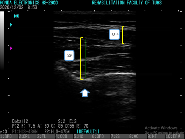
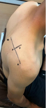
Musculoskeletal ultrasound imaging (USI), which has been shown to be a reliable method for measuring muscle and tendon thickness, was used to assess supraspinatus thickness . The approach used for probe placement closely followed the method described by Juul-Kristensen et al. The patient sat on a chair with the shoulder in a neutral position and the elbow fully extended. Using a linear transducer (5-12 MHz, HS-2600, Honda Electronics Co., Japan), the examiner identified the scapular spine by palpation and drew a line extending from its lateral to medial end (AM line) (Figure 1). The exact location of the probe, referred to as the P point, was identified at the midpoint of this line . The probe was held parallel to the supraspinatus fibers, ensuring the scapular notch was visible on the USI monitor (Figure 2). The maximum thickness of the supraspinatus muscle was identified and recorded at this location .
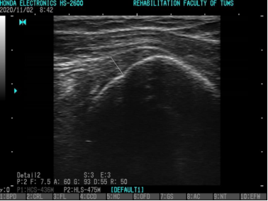
For the Supraspinatus tendon thickness measurement, the patient remained seated upright with both feet resting on the floor. She was instructed to put the tested hand on the ipsilateral iliac crest, with the elbow pointing backward (modified Crass position). The ultrasound transducer was positioned on the anterior aspect of the acromion in the coronal plane, where the greater tuberosity's footprint and superior facet were visible and the ultrasound image is shown in Figure 3 . The imaging process was repeated three times, and the average thickness was calculated . Echogenicity was determined from the pixel intensity of the ultrasound images using ImageJ software (National Institutes of Health, Bethesda, MD, USA) .
Figure 1. AM line, P point.
Figure 2. The exact placement of the probe to determine supraspinatus thickness, White Arrow shows Scapular Notch. SS: Supraspinatus, UT: Upper Trapezius.
Figure 3. Supraspinatus tendon thickness.
Pain pressure threshold
Patient's pain pressure threshold (PPT) was evaluated with an algometer during both the initial and final treatment sessions. The pressure (in newtons) was applied on the main bulk of the supraspinatus and the middle of the deltoid muscle, with a rate of 1kgf/s .
weight for exercise
The weight used in our treatment for low-load exercises was 20-40% 1 RM. American College of Sports Medicine (ACSM) defines 1RM as the maximum amount of weight that an individual can lift once through the full range of motion . Because directly determining 1RM may increase pain or aggravate inflammation in individuals with musculoskeletal injuries, 1RM was estimated using the Brzycki equation rather than being measured directly. In the first two weeks, the weight was set at 15-20% of 1RM, and for the subsequent two weeks, it was adjusted to 30-40% for the treatment protocol.
Brzycki → W/(1.0278-0.0278R)
W= weight lifted until failure
R= number of repetitions completed until failure
According to the prescribed formula, the patient was instructed to utilize a lightweight (1-2 kilograms) for performing scaption within a pain-free range of motion until muscular fatigue prevented further movement of the weight (repetition to failure).
2.3. Intervention
Before starting the intervention, the patient was screened for BFR contradictions including a thorough evaluation for any previous blood clot, blood pressure exceeding 180/100, acute infection, cancer, stroke, deep vein thrombosis (DVT), atrial fibrillation, and peripheral vascular disorders, and no contradiction was observed . Then, the strength of supraspinatus and scapular stabilizers including serratus anterior, lower and middle trapezius was measured using a dynamometer (MIE, Medical reaseach Itd, England). Additionally, ultrasound imaging (HS-2600 by Honda Electronics Co, Japan) was employed to assess the supraspinatus thickness, and algometry (Force-Gauge, 5020-Lutron, Taiwan) was utilized to determine the pain pressure threshold on the supraspinatus and deltoid muscles.
Following the baseline assessment, the patient adhered to a 4-week exercise therapy plan (3 times per week), as outlined in the table below. Pain intensity was recorded using the numeric pain rating scale (NPRS) 15 minutes before and after each treatment session. The intervention consisted of the following components (exercise details are presented in Table 1):
The exercise plan for scapular stabilizers (Table 1) which aims to balance the activity ratios of the upper trapezius to the lower trapezius, middle trapezius, and serratus anterior. This ratio should be the same or a bit less, as high ratios can lead to an imbalance in scapular stabilizer muscle activity. This imbalance may cause scapular dyskinesia, which can increase the risk of shoulder disorders, including rotator cuff tendinopathy .
Exercises designed to elicit maximum activation of the supraspinatus and infraspinatus muscles, including external rotation from 0 degrees abduction with a flexed elbow to 90 degrees, empty can, and full can .
Lower-limb aerobic exercise was incorporated to improve the function of the kinetic chain associated with scapular movement and to promote regional blood flow. Backward walking was chosen because it may reduce loading on the knee joint , while increasing activation of the core musculature involved in scapular control, making it a suitable exercise for this patient .
The exercise plan and the exercises are summarized in Table 1.
Table 1. The exercise plan and prescription.

Type of exercises

Intensity

Frequency

Protocol of BFR training

Name of Exercises

Scapular stabilization

20-40% 1RM

3 times per week

Rep:30/15/15/15 LOP: 50% Rest between sets: 30-60 s

scaption /Robbery

Rotator cuff strengthening

20-40% 1RM

3 times per week

Rep:30/15/15/15 Rest between sets: 30-60 s

External rotation with no abduction (while standing) full can

Aerobic

speed 1-2 m/h

3 times per week

10 minutes LOP:80 mmHg

Backward walking

Based on previously published BFR strengthening protocols, the patient performed four sets of each exercise, consisting of 30 repetitions in the first set followed by three sets of 15 repetitions. A 30-second rest period was provided between sets, while cuff pressure was maintained throughout the exercise using the continuous occlusion method . The intervention included 12 supervised treatment sessions over four weeks (three sessions per week). During each exercise, the patient reported the level of effort using the Rating of Perceived Exertion (RPE) scale, with the target intensity maintained between 6 and 8 (moderate to hard) . During the initial treatment session, the estimated 1RM, calculated using the Brzycki equation, was 2 kg. Exercise intensity was prescribed at 15-20% of the estimated 1RM during the first two weeks and was increased to 30-40% during the final two weeks of the intervention.
limb occlusion pressure (LOP)
To determine the LOP, a manual doppler (8 Mhz, EDAN doppler) was employed. OP was defined as the minimum cuff pressure required to completely eliminate the arterial pulse signal detected by the Doppler. According to current BFR exercise guidelines, cuff pressure corresponding to 50% of LOP for the upper limb and 80% of LOP for the lower limb is recommended during exercise . The BFR system used in this study was a Smart Cuff BFR device equipped with a portable vascular Doppler (EDAN Vascular Doppler, 8 MHz), medium cuffs (17.5-23 inches) for the lower extremities, and a small cuff (17 inches) for the upper extremity.
To determine LOP, the patient first remained seated in a relaxed position for 10 minutes. The cuff was then placed around the most proximal portion of the limb being tested. For the upper limb, the Doppler probe was positioned over the radial artery at approximately a 45° angle while avoiding excessive pressure that could interfere with blood flow. Cuff pressure was gradually increased until the arterial pulse signal disappeared, and this pressure was recorded as the LOP. The measured LOP values were 120 mmHg for the right upper limb, 240 mmHg for the right lower limb, and 160 mmHg for the left lower limb. To reduce the possibility of peripheral nerve compression, the upper-limb cuff was always positioned as proximally as possible.
Table 2 summarizes the LOP values and their progression throughout the intervention, which was adjusted to reduce discomfort and delayed-onset muscle soreness (DOMS). The table also presents the progression of treadmill speed during the backward-walking exercise program.
Table 2. Progression of LOP, weight and speed.

Second two weeks

First two weeks

Progression

30-40% 1RM

15-20% 1RM

Weight (% 1RM) (Kg)

50% LOP

30-40% LOP

LOP in upper limb (mmHg)

80% LOP

60-70% LOP

LOP in lower limb (mmHg)

1.5-2 mph

0.8-1 mph

Speed (mhp)

3. Results
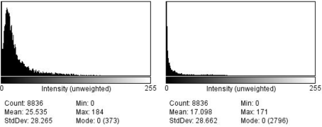
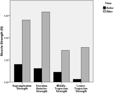
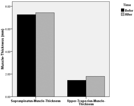
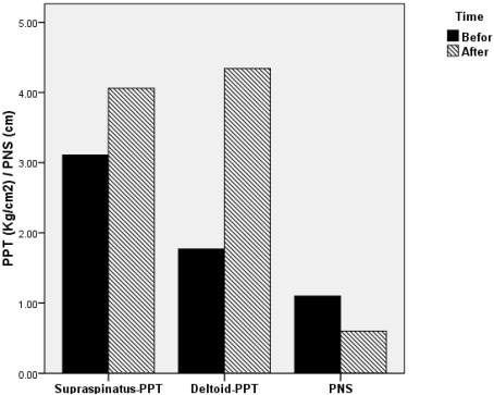
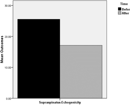
Table 3 compares the mean values of all outcome measures obtained at baseline and after the final treatment session, together with the patient's pain scores recorded using the NPRS. Following the 12-session intervention, increases were observed in the strength of the supraspinatus, serratus anterior, and the middle and lower trapezius muscles. Muscle thickness also increased in both the supraspinatus and upper trapezius, with a greater increase observed in the upper trapezius. The mean pain pressure threshold of both the supraspinatus and deltoid muscles improved after the intervention. In addition, the average NPRS score recorded 15 minutes before and after each treatment session decreased from 2.91 to 1.50 (Tables 3 and 4). A reduction in supraspinatus muscle echogenicity was also observed (Figure 4). Overall, favorable changes were found across all measured outcomes.
Figure 5 illustrates a comparison of muscle strength before and after treatment, while Figure 6 displays changes in muscle thickness. In the next figure (Figure 7), the comparison of the PPT of the deltoid and supraspinatus muscles, along with the NPRS are presented. Figure 8 specifically highlights the reduction in supraspinatus muscle echogenicity.
Table 3. Comparing the mean of outcome variables.

Outcomes

Before

After

Change (%Δ)

First session (mean)

Final session (mean)

Serratus anterior strength (N)

1.23

6.33

414.63

Middle trapezius strength (N)

0.90

2.86

217.77

Lower trapezius strength (N)

0.26

3.13

1103.84

PPT supraspinatus (Kg/cm2)

3.11

4.06

30.54

PPT deltoid (Kg/cm2)

1.77

4.34

145.19

Supraspinatus thickness (mm)

7.262

7.432

2.340

Echogenicity

25.535

17.098

-33.04

Upper trapezius thickness (mm)

1.450

1.798

24

Upper + supra thickness (mm)

9.040

9.379

3.75

Supraspinatus strength (N)

1.60

5.60

250

Supraspinatus tendon thickness (mm)

2.557

2.436

-0.047

NPRS

1.1

0.6

-45.45

FSS

4.33

2.33

-46.18

Figure 4. Echogenicity of supraspinatus muscle.
Figure 5. Muscle-Strength.
Figure 6. Muscle -Thickness.
Figure 7. Pain -Threshold.
Figure 8. Supraspinatus-Muscle -Echogenicity.
Table 4. Amount of NPRS before and after each session.

Number of the session

NPRS before the session

NPRS after the session

1

5

2

2

3

1

3

4

4

4

3

1

5

6

3

6

3

2

7

2

3

8

4

1

9

2

0

10

1

0

11

2

1

12

0

0

Mean

2.91

1.50

4. Discussion
In this case study, a patient diagnosed with MS underwent a novel treatment approach involving LLRE+BFR for supraspinatus tendinopathy. Substantial positive outcomes were observed, with notable improvements in supraspinatus and upper trapezius thickness, enhanced muscle strength in supraspinatus, serratus anterior, middle and lower trapezius. Furthermore, the treatment positively impacted PPT measurements for both deltoid and supraspinatus muscles, along with beneficial changes in NPRS parameters. Supraspinatus echogenicity also improved after intervention. Higher echogenicity of muscles is related to muscle weakness and atrophy. In other words, increased echogenicity could be interpreted as increased edema/inflammation, and decreased muscle quality and muscle glycogen. However, decreased echogenicity, which was the case in this study, indicates the exact opposite .
Most studies tend to focus on the distal effects of BFR, specifically emphasizing its influence on muscle strength and hypertrophy in muscles located below the cuff, while there are fewer studies investigating its proximal and contralateral effects, most of which report positive outcomes following BFR exercises . Studies by Bowmann and Lambert demonstrated that LLRE combined with BFR can improve the strength and muscle mass of shoulder muscles located proximal to the cuff. These findings indicate that this training approach may be useful in the rehabilitation of conditions such as shoulder tendinopathy, scapular dyskinesis, and shoulder instability pre- or post-surgery . In a study by Wang et al. (2026), BFR training yielded significant improvements in muscle strength and physical performance in different neurological populations, suggesting that the mechanisms responsible for muscle adaptation are preserved even in individuals with neurological impairment. Likewise, Blázquez-Fernández et al. (2024) showed clinically meaningful improvements in muscle strength and function with low-load BFR exercise among MS .
The findings of our study align with those of Bowmann et al. (2020) and Brumitt et al (2020) studies. Bowmann et al. found that hat LLRE+BFR increased proximal muscle strength and hypertrophy in healthy participants . Similarly, Brumitt et al reported that both LLRE+BFR and LLRE independently can improve strength and thickness of supraspinatus in healthy subjects . Consistent with these findings, the results of our investigation demonstrated increased muscle strength and hypertrophy after completing the LLRE+BFR program despite having supraspinatus tendinopathy. Following intervention, she achieved full range of motion in abduction and flexion without any signs of weakness.
Numerous studies have explored the effects of exercise with BFR on pain in both healthy individuals and individuals with musculoskeletal disorders . These studies report that using LLRE+BFR holds the potential to enhance muscle strength and hypertrophy, and to alleviate pain. Supraspinatus tendinopathy causes referral pain to the deltoid region and arm, and contributes to central sensitization in these patients. The primary source of pain in these cases is frequently attributed to an increase in central sensitization rather than tissue injuries . The findings of this study align with research conducted by Hughes et al. (2020) investigating the distal effect of BFR on pain perception. This study also demonstrated that LLRE+BFR can effectively alleviate proximal and distal pain in shoulder disorders . After treatment, the patient no longer experienced pain at rest and achieved full range of motion in active movements of the right arm. She also no longer suffered from sleep disturbances caused by shoulder pain.
In a study by Hughes et al. (2019) four possible neurophysiological mechanisms were introduced to elucidate the neurophysiological mechanisms underlying BFR's impact on pain alleviation . Firstly, BFR can stimulate baroreceptors in vessels, activating regions of the Central Nervous System (CNS) responsible for pressure and pulse control. This stimulation triggers descending inhibitory mechanisms, leading to pain relief. Second, according to the gate control theory, the temporary discomfort produced during BFR exercise may inhibit the perception of musculoskeletal pain. The third pathway is restricted venous return promotes the accumulation of metabolic by-products around the working muscles, stimulating type III and IV metaboreceptors. Activation of these receptors is believed to increase the release of endogenous opioids and endocannabinoids, contributing to pain relief. Finally, the reduction in blood flow accelerates fatigue of slow-twitch muscle fibers, leading to greater recruitment of fast-twitch fibers. Increased activation of these fibers may suppress pain transmission. Regarding this mechanism, training with high-intensity can effectively reduce pain. Despite the low-load nature of exercise with BFR, its addition substantially elevates exercise intensity and pressure, potentially leading to significant pain reduction.
Several studies have been conducted about the effects of BFR training in neurological rehabilitation. Recent systematic reviews have strengthened the evidence supporting BFR exercise in people with MS and other neurological disorders. Blázquez-Fernández et al. (2024) reported improvements in muscle strength, physical performance, fatigue, and exercise feasibility in individuals with MS after BFR training. Similarly, Jønsson et al. (2024) concluded that BFR exercise with proper protocols is generally safe and effective across neurological conditions. Furthermore, the most recent meta-analysis by Wang et al. (2026) supports BFR effectiveness on on strength and function in neurological disorders, including studies on MS patients . In a case report, Cohen et al. (2021) reported that LLRE+BFR in a 54-year-old patient with primary progressive MS resulted in improvements in the Multiple Sclerosis Walking Scale (MSWS-12), the Fatigue Severity Scale (FSS), patient-specific functional goals assessed by the Patient-Specific Functional Scale (PSFS)—including running for exercise and recreation for 45 minutes, climbing a flight of stairs with complete confidence, and driving safely without restrictions—as well as all 14 lower-extremity strength measures . Similarly, study conducted by Freitas et al (2021) demonstrated that MS patients can tolerate LLRE+BFR and require less muscular exertion compared to HLRE. Furthermore, this approach does not induce pain during exercises and DOMS after the intervention . These findings collectively suggest that training with BFR may potentially enhance muscle strength, size, and quality with reduced physiological stress comparing to traditional resistance training methods. This study revealed that the mean NPRS decreased 1.41 which is clinically significant (more than 1.39 ± 1.05) . Furthermore, at the baseline, the FSS score was 4.33, indicating severe MS-related fatigue. After treatment, the score decreased to 2.33, reflecting a 2-point improvement which is a clinically important change (>1.9 points) . This suggests that the prescribed exercises effectively reduced fatigue levels in the patient.
In our study, after employing LLRE+BFR, we observed a decrease in supraspinatus tendon thickness. Similarly, Skovlund and Cuddeford (2020) demonstrated that therapeutic exercise with BFR can reduce neovascularization and tendon thickness in tendinopathy, which results in pain reduction and improved tendon health . On the other hand, Brumitt et al. (2020) found that supraspinatus tendon CSA increased similarly with both low-load resistance training with BFR (LL-BFR) and without BFR (LL-RT), indicating that LL-BFR is not a proper option for the supraspinatus tendon. He suggests that the physiological effects observed in lower limb tendons may not translate to upper limb tendons . Furthermore, In some other studies an increase of tendon thickness is observed in association with LLRE+BFR. These mixed findings highlight the complexity of tendon adaptations and underscore the need for further research to fully understand the implications and underlying mechanisms of tendon thickness changes, particularly in specific populations like MS patients.
The physiological mechanisms responsible for the increases in muscle strength and hypertrophy following LLRE+BFR have not been fully clarified, although several explanations have been proposed . Because the mechanical load during LLRE+BFR is relatively low, it is believed that metabolic stress, rather than mechanical tension, is the primary stimulus for muscle adaptation . During LLRE+BFR, muscle ischemia generated during the exercises results in metabolic stress on muscle . This condition promotes the accumulation of metabolites, including lactate, inside the muscle cells, which increases osmotic pressure by drawing interstitial fluid into the cells . Consequently, this accumulation leads to inflammation within the muscle cell, activating protein generation pathways such as mTOR and MAPK . The resulting cell swelling activates intracellular signaling pathways involved in protein synthesis, such as the mTOR and MAPK pathways, ultimately promoting muscle hypertrophy . Therefore, metabolic stress is considered one of the principal mechanisms underlying the anabolic effects of BFR , leading to improvements in muscle strength and hypertrophy in muscles located distal to the cuff.
The mechanism responsible for the proximal effects of BFR is less well established. One possible explanation is that fatigue of the distal muscles during LLRE+BFR increases the recruitment and compensatory activation of proximal muscles. In addition, the biceps and triceps muscles, which are located distal to the cuff and are more responsive to strength and hypertrophy adaptations, contribute to glenohumeral stability. Their increased activation may indirectly facilitate improvements in the strength of muscles located proximal to the cuff.
To the best of our knowledge, no previous study has investigated the proximal effects of LLRE+BFR in patients with multiple sclerosis who present with shoulder pain. Our positive findings underscore the necessity for further investigation and a more comprehensive exploration into the effectiveness of LLRE+BFR for MS patients with musculoskeletal disorders.
5. Conclusion
This study suggests that LLRE+BFR could be a potentially effective option for MS patients with supraspinatus tendinopathy. After the 4-week intervention, improvements were observed in strength, muscle and tendon thickness, pain, and fatigue with no adverse events. These findings support the potential proximal effects of LLRE+BFR on the shoulder and indicate that this approach may be a viable alternative to HLRE for those who cannot tolerate high loads. However, as this report describes a single patient, the findings should be interpreted with caution and cannot be generalized.
6. Recommendations
LLRE+BFR could be considered an adjunct rehabilitation strategy in clinics for MS patients with concomitant shoulder tendinopathy, especially when fatigue and pain limit their ability to tolerate high loads. There is a need for studies with larger sample sizes, longer follow-up periods and controlled study designs to confirm the effectiveness of this approach.
Abbreviations

HLRE

High-Load Resistance Exercise

BFR

Blood Flow Restriction

LLRE

Low-Load Resistance Exercise

LLRE+BFR

Low-Load Resistance Exercise Combined with Blood Flow Restriction

RM

Repetition Maximum

MS

Multiple Sclerosis

FSS

Fatigue Severity Scale

MRI

Magnetic Resonance Imaging

MMT

Manual Muscle Testing

USI

Musculoskeletal Ultrasound Imaging

PPT

Pain Pressure Threshold

ACSM

American College of Sports Medicine

NPRS

Numeric Pain Rating Scale

RPE

Rating of Perceived Exertion

LOP

Limb Occlusion Pressure

DOMS

Delayed-Onset Muscle Soreness

DVT

Deep Vein Thrombosis

CSA

Cross-Sectional Area

CNS

Central Nervous System

MSWS-12

Multiple Sclerosis Walking Scale-12

PSFS

Patient-Specific Functional Scale

mTOR

Mammalian Target of Rapamycin

MAPK

Mitogen-Activated Protein Kinase

Author Contributions
Zahra Poursaleh Begi: Conceptualization, Methodology, Resources, Supervision
Bahram Tabatabaei: Investigation, Writing – original draft, Visualization
Nastaran Bahadorani: Data curation, Methodology, Writing – review & editing
Conflicts of Interest
The authors declare no conflicts of interest.
References
[1] Luime, J., Koes, B., Hendriksen, I., Burdorf, A., Verhagen, A., Miedema, H., & Verhaar, J. (2004). Prevalence and incidence of shoulder pain in the general population; a systematic review. Scandinavian Journal of Rheumatology, 33(2), 73–81.
[2] Oliva F, Piccirilli E, Bossa M, et al. I. S. Mu. L. T - Rotator Cuff Tears Guidelines. Muscles Ligaments Tendons J. 2016; 5(4): 227-263. Published 2016 Feb 13.
[3] Heron SR, Woby SR, Thompson DP. Comparison of three types of exercise in the treatment of rotator cuff tendinopathy/shoulder impingement syndrome: A randomized controlled trial. Physiotherapy. 2017; 103(2): 167-173.
[4] Lewis J, McCreesh K, Roy JS, Ginn K. Rotator Cuff Tendinopathy: Navigating the Diagnosis-Management Conundrum. J Orthop Sports Phys Ther. 2015; 45(11): 923-937.
[5] Grønfeldt BM, Lindberg Nielsen J, Mieritz RM, Lund H, Aagaard P. Effect of blood-flow restricted vs heavy-load strength training on muscle strength: Systematic review and meta-analysis. Scand J Med Sci Sports. 2020; 30(5): 837-848.
[6] Cohen ET, Cleffi N, Ingersoll M, Karpatkin HI. Blood-Flow Restriction Training for a Person With Primary Progressive Multiple Sclerosis: A Case Report. Phys Ther. 2021; 101(3): pzaa224.
[7] Garber CE, Blissmer B, Deschenes MR, et al. American College of Sports Medicine position stand. Quantity and quality of exercise for developing and maintaining cardiorespiratory, musculoskeletal, and neuromotor fitness in apparently healthy adults: guidance for prescribing exercise. Med Sci Sports Exerc. 2011; 43(7): 1334-1359.
[8] Sato, Y.. (2005). The history and future of KAATSU Training. International Journal of KAATSU Training Research. 1. 1-5.
[9] Lixandrão ME, Ugrinowitsch C, Berton R, et al. Magnitude of Muscle Strength and Mass Adaptations Between High-Load Resistance Training Versus Low-Load Resistance Training Associated with Blood-Flow Restriction: A Systematic Review and Meta-Analysis. Sports Med. 2018; 48(2): 361-378.
[10] Hughes L, Paton B, Rosenblatt B, Gissane C, Patterson SD. Blood flow restriction training in clinical musculoskeletal rehabilitation: a systematic review and meta-analysis. Br J Sports Med. 2017; 51(13): 1003-1011.
[11] Patterson SD, Hughes L, Warmington S, et al. Blood Flow Restriction Exercise: Considerations of Methodology, Application, and Safety. Front Physiol. 2019; 10: 533. Published 2019 May 15.
[12] Hughes L, Patterson SD. The effect of blood flow restriction exercise on exercise-induced hypoalgesia and endogenous opioid and endocannabinoid mechanisms of pain modulation. J Appl Physiol (1985). 2020; 128(4): 914-924.
[13] Jørgensen SL, Kierkegaard-Brøchner S, Bohn MB, Høgsholt M, Aagaard P, Mechlenburg I. Effects of blood-flow restricted exercise versus conventional resistance training in musculoskeletal disorders-a systematic review and meta-analysis. BMC Sports Sci Med Rehabil. 2023; 15(1): 141. Published 2023 Oct 25.
[14] Cruickshank TM, Reyes AR, Ziman MR. A systematic review and meta-analysis of strength training in individuals with multiple sclerosis or Parkinson disease. Medicine (Baltimore). 2015; 94(4): e411.
[15] Bjarnadottir OH, Konradsdottir AD, Reynisdottir K, Olafsson E. Multiple sclerosis and brief moderate exercise. A randomised study. Mult Scler. 2007; 13(6): 776-782.
[16] Severijns D, Zijdewind I, Dalgas U, Lamers I, Lismont C, Feys P. The Assessment of Motor Fatigability in Persons With Multiple Sclerosis: A Systematic Review. Neurorehabil Neural Repair. 2017; 31(5): 413-431.
[17] Smith RM, Adeney-Steel M, Fulcher G, Longley WA. Symptom change with exercise is a temporary phenomenon for people with multiple sclerosis. Arch Phys Med Rehabil. 2006; 87(5): 723-727.
[18] Bowman EN, Elshaar R, Milligan H, et al. Upper-extremity blood flow restriction: the proximal, distal, and contralateral effects-a randomized controlled trial. J Shoulder Elbow Surg. 2020; 29(6): 1267-1274.
[19] Gismervik SØ, Drogset JO, Granviken F, Rø M, Leivseth G. Physical examination tests of the shoulder: a systematic review and meta-analysis of diagnostic test performance. BMC Musculoskelet Disord. 2017; 18(1): 41. Published 2017 Jan 25.
[20] Jancuska J, Matthews J, Miller T, Kluczynski MA, Bisson LJ. A Systematic Summary of Systematic Reviews on the Topic of the Rotator Cuff. Orthop J Sports Med. 2018; 6(9): 2325967118797891. Published 2018 Sep 21.
[21] Nakajima, T. & Morita, T. & Sato, Y.. (2011). Key considerations when conducting KAATSU training. International Journal of KAATSU Training Research. 7. 1-6.
[22] Kacin, Alan & Rosenblatt, Ben & Tomc Zargi, Tina & Biswas, Anita. (2016). SAFETY CONSIDERATIONS WITH BLOOD FLOW RESTRICTED RESISTANCE TRAINING. Annales Kinesiologiae. vol 6. 3-26.
[23] Hannah DC, Scibek JS, Carcia CR. STRENGTH PROFILES IN HEALTHY INDIVIDUALS WITH AND WITHOUT SCAPULAR DYSKINESIS. Int J Sports Phys Ther. 2017; 12(3): 305-313.
[24] Robertson JA. F. P. Kendall and E. K. McCreary “Muscles, Testing and Function” (Third Edition). Br J Sports Med. 1984 Mar; 18(1): 25.
[25] Krzesniak-Swinarska M, Caress JB, Cartwright MS. Neuromuscular ultrasound for evaluation of scapular winging. Muscle Nerve. 2017; 56(1): 7-14.
[26] Juul-Kristensen B, Bojsen-Møller F, Holst E, Ekdahl C. Comparison of muscle sizes and moment arms of two rotator cuff muscles measured by ultrasonography and magnetic resonance imaging. Eur J Ultrasound. 2000; 11(3): 161-173.
[27] Papatzika F, Papandreou M, Ekizos A, Panteli C, Arampatzis A. Reliability and Limits of Agreement of the Supraspinatus Muscle Anatomical Cross-Sectional Area Assessment by Ultrasonography. Ultrasound Med Biol. 2015; 41(7): 1821-1826.
[28] Ishigaki T, Yoshino K, Hirokawa M, Sugawara M, Yamanaka M. Supraspinatus tendon thickness and subacromial impingement characteristics in younger and older adults. BMC Musculoskelet Disord. 2022; 23(1): 234. Published 2022 Mar 11.
[29] Wong V, Spitz RW, Bell ZW, et al. Exercise induced changes in echo intensity within the muscle: a brief review. J Ultrasound. 2020; 23(4): 457-472.
[30] Wong V, Abe T, Chatakondi RN, et al. The influence of biological sex and cuff width on muscle swelling, echo intensity, and the fatigue response to blood flow restricted exercise. J Sports Sci. 2019; 37(16): 1865-1873.
[31] Medicine AC of S, Brubaker PH, Otto RM, Armstrong LE. ACSM’s Guidelines For Exercise Testing And Prescription. 7th edition. Whaley MH, editor. Baltimore: Lippincott Williams & Wilkins; 2006.
[32] McNair PJ, Colvin M, Reid D. Predicting maximal strength of quadriceps from submaximal performance in individuals with knee joint osteoarthritis. Arthritis Care Res (Hoboken). 2011; 63(2): 216-222.
[33] Schory A, Bidinger E, Wolf J, Murray L. A SYSTEMATIC REVIEW OF THE EXERCISES THAT PRODUCE OPTIMAL MUSCLE RATIOS OF THE SCAPULAR STABILIZERS IN NORMAL SHOULDERS. Int J Sports Phys Ther. 2016; 11(3): 321-336.
[34] Ganderton, Charlotte & Pizzari, Tania. (2013). A Systematic Literature Review of the Resistance Exercises That Promote Maximal Muscle Activity of the Rotator Cuff in Normal Shoulders. Shoulder & Elbow. 5. 120-135.
[35] Brumitt J, Hutchison MK, Kang D, et al. Blood Flow Restriction Training for the Rotator Cuff: A Randomized Controlled Trial. Int J Sports Physiol Perform. 2020; 15(8): 1175-1180. Published 2020 Aug 19.
[36] Roos PE, Barton N, van Deursen RW. Patellofemoral joint compression forces in backward and forward running. J Biomech. 2012; 45(9): 1656-1660.
[37] Richardson E, Lewis JS, Gibson J, et al. Role of the kinetic chain in shoulder rehabilitation: does incorporating the trunk and lower limb into shoulder exercise regimes influence shoulder muscle recruitment patterns? Systematic review of electromyography studies. BMJ Open Sport Exerc Med. 2020; 6(1): e000683. Published 2020 Apr 22.
[38] Bowman EN, Elshaar R, Milligan H, et al. Proximal, Distal, and Contralateral Effects of Blood Flow Restriction Training on the Lower Extremities: A Randomized Controlled Trial. Sports Health. 2019; 11(2): 149-156.
[39] Lambert, Bradley & Hedt, Corbin & Epner, Eden & Chaliki, Kalyan & Wang, Christine & Lee, Joshua & Rossano, Aya & Moreno, Michael & McCulloch, Patrick. (2019). B. F. R. For Proximal Benefit: Blood Flow Restriction Therapy For The Shoulder?
[40] Plinsinga ML, Brink MS, Vicenzino B, van Wilgen CP. Evidence of Nervous System Sensitization in Commonly Presenting and Persistent Painful Tendinopathies: A Systematic Review. J Orthop Sports Phys Ther. 2015; 45(11): 864-875.
[41] Freitas, Eduardo & Miller, Ryan & Heishman, Aaron & Aniceto, Rodrigo & Larson, Rebecca & Pereira, Hugo & Bemben, Debra & Bemben, Michael. (2020). The perceptual responses of individuals with multiple sclerosis to blood flow restriction versus traditional resistance exercise. Physiology & Behavior. 229. 113219.
[42] Kendrick DB, Strout TD. The minimum clinically significant difference in patient-assigned numeric scores for pain. Am J Emerg Med. 2005; 23(7): 828-832.
[43] Learmonth YC, Dlugonski D, Pilutti LA, Sandroff BM, Klaren R, Motl RW. Psychometric properties of the Fatigue Severity Scale and the Modified Fatigue Impact Scale. J Neurol Sci. 2013; 331(1-2): 102-107.
[44] Cuddeford T, Brumitt J. IN-SEASON REHABILITATION PROGRAM USING BLOOD FLOW RESTRICTION THERAPY FOR TWO DECATHLETES WITH PATELLAR TENDINOPATHY: A CASE REPORT. Int J Sports Phys Ther. 2020; 15(6): 1184-1195.
[45] Skovlund, S., Aagaard, P., Larsen, P. T., Svensson, R. B., Kjaer, M., Magnusson, S. P., & Couppé, C. (2020). The effect of low-load resistance training with blood flow restriction on chronic patellar tendinopathy – a case series. Translational Sports Medicine, 3(4), 342-352.
[46] Arampatzis A, Karamanidis K, Albracht K. Adaptational responses of the human Achilles tendon by modulation of the applied cyclic strain magnitude. J Exp Biol. 2007; 210(Pt 15): 2743-2753.
[47] Docking SI, Cook J. How do tendons adapt? Going beyond tissue responses to understand positive adaptation and pathology development: A narrative review. J Musculoskelet Neuronal Interact. 2019; 19(3): 300-310.
[48] Ferguson RA, Hunt JEA, Lewis MP, et al. The acute angiogenic signalling response to low-load resistance exercise with blood flow restriction. Eur J Sport Sci. 2018; 18(3): 397-406.
[49] Takada S, Okita K, Suga T, et al. Low-intensity exercise can increase muscle mass and strength proportionally to enhanced metabolic stress under ischemic conditions. J Appl Physiol (1985). 2012; 113(2): 199-205.
[50] Behringer M, Willberg C. Application of Blood Flow Restriction to Optimize Exercise Countermeasures for Human Space Flight. Front Physiol. 2019; 10: 33. Published 2019 Jan 25.
[51] Hughes L, Patterson SD. Low intensity blood flow restriction exercise: Rationale for a hypoalgesia effect. Med Hypotheses. 2019; 132: 109370.
[52] Blázquez-Fernández, A., Marcos-Antón, S., & Cano-de-la-Cuerda, R. (2024). Effectiveness and Feasibility of Blood Flow Restriction Training for People with Multiple Sclerosis: A Systematic Review. Neurology International, 16(6), 1385-1404.
[53] Jønsson AB, Krogh S, Laursen HS, Aagaard P, Kasch H, Nielsen JF. Safety and efficacy of blood flow restriction exercise in individuals with neurological disorders: A systematic review. Scand J Med Sci Sports. 2024; 34(1): e14561.
[54] Wang H, Cao Y, Zhang J, et al. Effectiveness of Blood Flow Restriction Training in Neurological Rehabilitation: A Systematic Review and Meta-analysis. Sports Med Open. 2026; 12(1): 53. Published 2026 May 6.
Cite This Article
  • APA Style

    Begi, Z. P., Tabatabaei, B., Bahadorani, N. (2026). Effects of Low-Load Exercises with Blood Flow Restriction on Shoulder of a Multiple Sclerosis Patient with Shoulder Tendinopathy: A Case Study. International Journal of Sports Science and Physical Education, 11(2), 32-44. https://doi.org/10.11648/j.ijsspe.20261102.12

    Copy | Download

    ACS Style

    Begi, Z. P.; Tabatabaei, B.; Bahadorani, N. Effects of Low-Load Exercises with Blood Flow Restriction on Shoulder of a Multiple Sclerosis Patient with Shoulder Tendinopathy: A Case Study. Int. J. Sports Sci. Phys. Educ. 2026, 11(2), 32-44. doi: 10.11648/j.ijsspe.20261102.12

    Copy | Download

    AMA Style

    Begi ZP, Tabatabaei B, Bahadorani N. Effects of Low-Load Exercises with Blood Flow Restriction on Shoulder of a Multiple Sclerosis Patient with Shoulder Tendinopathy: A Case Study. Int J Sports Sci Phys Educ. 2026;11(2):32-44. doi: 10.11648/j.ijsspe.20261102.12

    Copy | Download

  • @article{10.11648/j.ijsspe.20261102.12,
      author = {Zahra Poursaleh Begi and Bahram Tabatabaei and Nastaran Bahadorani},
      title = {Effects of Low-Load Exercises with Blood Flow Restriction on Shoulder of a Multiple Sclerosis Patient with Shoulder Tendinopathy: A Case Study},
      journal = {International Journal of Sports Science and Physical Education},
      volume = {11},
      number = {2},
      pages = {32-44},
      doi = {10.11648/j.ijsspe.20261102.12},
      url = {https://doi.org/10.11648/j.ijsspe.20261102.12},
      eprint = {https://article.sciencepublishinggroup.com/pdf/10.11648.j.ijsspe.20261102.12},
      abstract = {Blood flow restriction (BFR) training involves placing an inflatable cuff around the proximal part of a limb to partially restrict arterial inflow during exercise. When combined with low-load resistance exercise (LLRE), this approach has been shown to produce improvements in muscle strength and hypertrophy comparable to those achieved with conventional high-load resistance training in healthy individuals. Through its proximal effects, LLRE combined with BFR may also enhance muscle strength and size in individuals with supraspinatus tendinopathy. Although high-load resistance training is effective for increasing muscle strength and hypertrophy, its application in people with multiple sclerosis (MS) is often limited because of fatigue. This case report describes the proximal effects of an LLRE+BFR program in a patient with MS and supraspinatus tendinopathy. Our case was a 48-year-old woman with a 5-year history of relapsing-remitting MS, who presented with right shoulder pain and muscle weakness in the right shoulder at rest and while elevation. She received a LLRE+BFR program for her right shoulder and aerobic exercises based on the BFR protocols in 12 sessions over 4 weeks. Outcome measures were collected before the intervention and after 4 weeks and included: 1) strength of supraspinatus and four scapular stabilizer muscles. 2) thickness of supraspinatus and upper trapezius muscles. 3) thickness of supraspinatus tendon 4) pain pressure threshold of supraspinatus and deltoid muscles. 5) Supraspinatus muscle echogenicity. Improvement was observed in the strength of supraspinatus and scapular stabilizers. Thickness of supraspinatus and upper trapezius muscles and pain pressure threshold in the supraspinatus and deltoid muscles also increased. Furthermore, supraspinatus echogenicity and tendon thickness decreased. The patient experienced a significant improvement in all outcomes following the use of a LLRE+BFR program. This program may be an option for strengthening training with less stress in MS people with supraspinatus tendinopathy.},
     year = {2026}
    }
    

    Copy | Download

  • TY  - JOUR
    T1  - Effects of Low-Load Exercises with Blood Flow Restriction on Shoulder of a Multiple Sclerosis Patient with Shoulder Tendinopathy: A Case Study
    AU  - Zahra Poursaleh Begi
    AU  - Bahram Tabatabaei
    AU  - Nastaran Bahadorani
    Y1  - 2026/08/17
    PY  - 2026
    N1  - https://doi.org/10.11648/j.ijsspe.20261102.12
    DO  - 10.11648/j.ijsspe.20261102.12
    T2  - International Journal of Sports Science and Physical Education
    JF  - International Journal of Sports Science and Physical Education
    JO  - International Journal of Sports Science and Physical Education
    SP  - 32
    EP  - 44
    PB  - Science Publishing Group
    SN  - 2575-1611
    UR  - https://doi.org/10.11648/j.ijsspe.20261102.12
    AB  - Blood flow restriction (BFR) training involves placing an inflatable cuff around the proximal part of a limb to partially restrict arterial inflow during exercise. When combined with low-load resistance exercise (LLRE), this approach has been shown to produce improvements in muscle strength and hypertrophy comparable to those achieved with conventional high-load resistance training in healthy individuals. Through its proximal effects, LLRE combined with BFR may also enhance muscle strength and size in individuals with supraspinatus tendinopathy. Although high-load resistance training is effective for increasing muscle strength and hypertrophy, its application in people with multiple sclerosis (MS) is often limited because of fatigue. This case report describes the proximal effects of an LLRE+BFR program in a patient with MS and supraspinatus tendinopathy. Our case was a 48-year-old woman with a 5-year history of relapsing-remitting MS, who presented with right shoulder pain and muscle weakness in the right shoulder at rest and while elevation. She received a LLRE+BFR program for her right shoulder and aerobic exercises based on the BFR protocols in 12 sessions over 4 weeks. Outcome measures were collected before the intervention and after 4 weeks and included: 1) strength of supraspinatus and four scapular stabilizer muscles. 2) thickness of supraspinatus and upper trapezius muscles. 3) thickness of supraspinatus tendon 4) pain pressure threshold of supraspinatus and deltoid muscles. 5) Supraspinatus muscle echogenicity. Improvement was observed in the strength of supraspinatus and scapular stabilizers. Thickness of supraspinatus and upper trapezius muscles and pain pressure threshold in the supraspinatus and deltoid muscles also increased. Furthermore, supraspinatus echogenicity and tendon thickness decreased. The patient experienced a significant improvement in all outcomes following the use of a LLRE+BFR program. This program may be an option for strengthening training with less stress in MS people with supraspinatus tendinopathy.
    VL  - 11
    IS  - 2
    ER  - 

    Copy | Download

Author Information
  • School of Rehabilitation, Tehran University of Medical Sciences (TUMS), Tehran, Iran
  • School of Rehabilitation, Tehran University of Medical Sciences (TUMS), Tehran, Iran

  • School of Rehabilitation, Tehran University of Medical Sciences (TUMS), Tehran, Iran